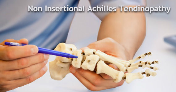
Non Insertional Achilles Tendinopathy
By Jonathan Larholt FCPodS, Consultant Podiatric Surgeon
Please get in Contact to book an appointment with Mr. Jonathan Larholt
Abstract
A fascinating article investigating the nature of Non-Insertional Achilles Tendinopathy, its causes, presentation, and common treatments. Written by Consultant Podiatric Surgeon Mr. Jonathan Larholt, FCPodS, this overview is a captivating insight into this degenerative condition and available treatments.
Article
The London marathon was the culmination of many months of training for some 40,000 people when they commenced the 26.2 mile run. Many months before the event the training began and over time the amount of miles that were run increased. With this increase in activity there is also the risk of injuring the Achilles tendon, which is a commonly affected structure.
The Achilles tendon is the largest tendon of the body and acts by lifting the heel off of the ground and transferring the weight / force to the ball of the foot, contributing to forward propulsion.
The Achilles tendon is situated at the back of the ankle and foot and the tendon is formed from two muscles, which are collectively termed the calf muscle. The tendon begins in the lower third of the leg and inserts into the back of the heel bone where it progresses to the underside of the heel to merge with the plantar fascia (a thick fibrous band on the sole of the foot which supports the arch and contributes to the function of the foot. Injury of the origin of the plantar fascia on the underside of the heel bone is the most common cause of heel pain, plantar fasciitis).
Unlike other tendons of the body the Achilles tendons fibres are orientated in a spiral manner; this allows the tendon to store energy for when the heel comes off of the ground, the “spring in your step!”
The Achilles tendon is a very powerful tendon and is prone to injury. Injury can occur in one of two areas; within the main body of the tendon, termed non-insertional Achilles tendinopathy or where it inserts onto the heel bone: insertional Achilles tendinopathy. This blog focuses on non-insertional Achilles tendinopathy.
Non-insertional Achilles tendinopathy develops when the force placed upon the tendon is greater than the tendons strength (tensile). This force is often repetitive, however, an isolated incident can occur to cause injury and often produce rupture of the tendon.
What may surprise people is that Achilles tendinopathy is not an inflammatory condition but a degenerative condition (The term tendinopathy: degenerative / tendinitis inflammatory). When the tendon becomes injured the damaged collagen begins to breakdown and releases chemicals which sensitise the nerves to produce pain.
Non-insertional Achilles tendinopathy is experienced as pain and swelling approximately 2 to 6 centimetres above the heel bone. The pain can be associated with swelling, morning stiffness or stiffness that can return after periods of rest, stiffness associated with activity, reduction in motion and pain on motion. A common factor of the condition is thickening of the tendon itself. As well as the tendon being affected the sheath around the tendon can become inflamed and if this is the case a creaking sensation and sound may be heard during activity, pain associated with the tendon sheath is inflammatory in nature.
There are many factors which can lead to non-insertional Achilles tendinopathy and these are often divided into extrinsic (things outside of the body) and intrinsic (associated with the body). Some of these factors are modifiable and can be addressed to reduce the risk of developing Achilles tendinopathy and assist in its management.
Extrinsic factors include:
- Running surface (modifiable): running on a hill or cambered surface can increase stress on the tendons
- Overuse / over training (modifiable): increases in the duration and or frequency of training. This leads to an imbalance with an increase in wear and insufficient time for repair.
- Inflexibility / improper stretching (modifiable): the tendons are tighter and more susceptible to tensile loading and injury.
- Footwear: worn out trainers or improper footwear (modifiable): If the heel of the shoe is worn out this would lead to altered foot mechanics
Intrinsic factors include:
- Increased age: (as we age our tendons become more inflexible and susceptible to injury)
- Obesity (modifiable): Increased weight produces increased load onto the tendon.
- Genetic predisposition: recently there has been much research into genetic markers and the development of Achilles tendinopathy
- Poor foot function (modifiable through orthosis/insoles): A pronated foot produces compensation within the foot and ankle and leads to a whipping action of the Achilles which can lead to damage.
- Malnutrition (modifiable): A poor diet does not provide the body with the building blocks to allow for the reparative process to take place
- Previous tendinopathy: Previous tendinopathy is a large risk factor for developing future tendinopathy
- Antibiotics (modifiable): The class of antibiotics Fluoroquinolones to include Cirpofloxacillin can reduce the strength of tendons and increase their susceptibility to injury
- Tendinopathy secondary to the plantaris tendon: The plantaris tendon is a long tendon on the inside aspect of the leg which is in close proximity to the Achilles tendon. It is absent in a third of the population and is somewhat redundant in humans but is important in jumping mammals such as Kangaroos. This tendon can produce pressure onto the Achilles tendon and lead to an Achilles tendinopathy
To further assess the pathology investigations such as ultrasound and MRI are often requested. Investigations also allow other pathologies such as tears to be ruled out. The management of a tear is very different from a tendinopathy.
The scans often show a variety of changes:
- Thickening of the tendon
- Blood vessel infiltration: accompanying the blood vessel can be nerves which can produce the pain associated with the condition.
- Degeneration
- Tears
Treatment involves a number of different approaches:
- Assessment of modifiable risk factors (outlined above)
- Loading of the tendon:
Isometric loading (no change in the length of the muscle):
stretching Isotonic loading (change in length but tension remains the same) to include eccentric stretching. This is where the heel is dropped beneath the level of a step, which puts tension on the muscle and tendon and helps to stimulate the repair process.
Other treatments include:
- Shockwave therapy: This is a machine that passes shockwaves to the affected area to stimulate repair and reduce pain.
- High Volume Injection Therapy: This involves using ultrasound to guide a needle between the tendon and tendon sheath followed by the administration of a large volume of saline. This separates the tendon sheath (paratenon) from the tendon itself and can reduce the amount of new blood vessels into the tendon, part of the pathology. With this treatment the pain also reduces and patients are able to perform their stretching exercises more appropriately.
- Sclerosing therapy: This is where a sclerosing agent is injected around areas of microtears. This can reduce pain and improve function, this treatment is also performed under ultrasound guidance.
- Plasma Rich Platelet (PRP): PRP is used to promote healing of the tendon and is obtained by drawing blood from the patient and spinning the blood to obtain certain products which are then injected around the tendon. The evidence for PRP is not as strong as the aforementioned treatments.
Prior to any surgical management conservative and medical care (outlined above) is continued for six months.
There are a number of different surgical approaches and these are dependent on the amount of tendon degeneration and associated pathologies e.g. inflammation of the tendon sheath or enlarged plantaris tendon.
Surgical management includes:
- Removal of degenerative areas and longitudinal incisions within the tendon to stimulate repair.
- Surgical stripping of the paratenon to reduce the infiltration of new blood vessels and allow for tendon healing.
- Resection of the plantaris tendon
- Tendon transfer. If the degeneration of the tendon is greater than 50% a tendon transfer is often required to reinforce the Achilles.
The outcome of surgical management can be variable and should only be entertained once all conservative care has failed. With all treatments a cure is not guaranteed, but patients are in less pain and more functional and thereby able to enhance their activities or daily living and quality of life.
This blog aims to provide you with an oversight of the condition and available treatments. Achilles tendinopathy can be a debilitating condition whether it is insertional or non-insertional and earlier treatment can prevent further injury and debilitation.


